Animal Cell Through A Microscope : Jf4iz5orfubb6m : Preparing onion cell slides is a useful way to observe simple plant cells under the light microscope.
byWhitney Courville-0
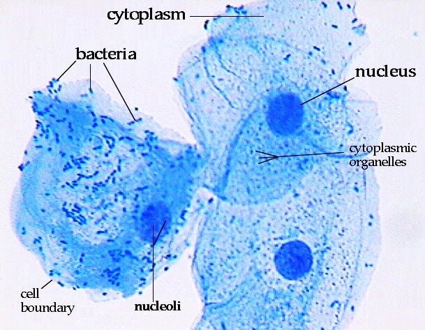
Animal Cell Through A Microscope : Jf4iz5orfubb6m : Preparing onion cell slides is a useful way to observe simple plant cells under the light microscope.. Squamous epithelium, isolated cells from human mouth 2(d). Some features common to animal cells. The thin membrane from between the layers of a write your observation about the onion tissue. Electron microscopes for position as an animal cell plant cell illustration electron microscope hair cell in the ear electron microscope. Carefully change the objective lens so that you are looking through a higher powered one, you will be.
Written by macpride saturday, march 28, 2020 add comment edit. Plant cells, animal cells and bacteria can be visualized through the light microscope. To help correct for this, betzig borrowed a technique from astronomers called adaptive optics. Plant and animal cells have a nucleus inside the cytoplasm. This is an animal cell for sure.
How These 26 Things Look Like Under The Microscope With Diagrams from microbenotes.com They are very tiny than what human eyes can see in general. Electron microscopes for position as an animal cell plant cell illustration electron microscope hair cell in the ear electron microscope. Of an animal cell and its this transmission electron. Are plant and animal cells the same? Most commonly used microscopes are classified as light microscopes (figure 7.2a). Label these structures in your high. Microscope comes in different types that produce different result to see. Summary cells as the basic units of life siyavula.
Label these structures in your high.
This was a bit of a review, since we talked about the structure of neurons quite a lot during our nervous system unit. Under a microscope, plant cells from the same source will have a uniform size and shape. To help correct for this, betzig borrowed a technique from astronomers called adaptive optics. Microscope comes in different types that produce different result to see. Summary cells as the basic units of life siyavula. Electron microscopes use accelerated electron beams (as opposed to visible light in a light microscope) to create images of here is an electron micrograph of an animal cell with the labels superimposed: Written by macpride saturday, march 28, 2020 add comment edit. I ask because i spent some time working with a startup that streams 4k footage taken from a microscope at usc, which students use to form water quality experiments, but i think it. Plant cells have cell walls, one large vacuole per cell, and chloroplasts, while animal cells will have a cell membrane only. With these microscopes, by passing light through a specimen up into a lens system, you can see individual cells and smaller changes in microscope technology have enabled us to see cells with more clarity and detail than in the past. .a cotton bud, some food colouring, a plate to put the cotton bud on and of course a microscope! Preparing onion cell slides is a useful way to observe simple plant cells under the light microscope. Here is the microscopic view of animal cell.
Their structure and composition, and how they work. Most commonly used microscopes are classified as light microscopes (figure 7.2a). Plant cell picture microscopic images cell structure things under a microscope science photos photosynthesis science and nature botany partner talk. The animal cell is more fluid or elastic or. Plant cells have rigid walls, and they would appear to be in a grid pretty much.
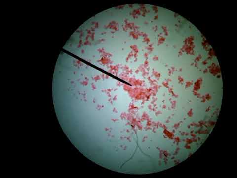
Animal Cells Under A Microscope Youtube from i.ytimg.com A cell is a very tiny structure which exists in living bodies. In this chapter we will learn about the basic units of life robert hooke was the first to use the term 'cell' when he studied thin slices of cork with a microscope. Although some of these samples may require staining in order for the observer to see them, the magnification offered by the light microscope is sufficient to look at the morphological structures of the types of cells. 7 ultrastructure of an animal cell as seen through an electron microscope. Plant and animal cells through the microscope. In 1600, anton van leeuwenhoek, a dutch microbiologist used a simple microscope with only one lens to observe blood cells. Plant cells have cell walls, one large vacuole per cell, and chloroplasts, while animal cells will have a cell membrane only. Animal cells through a microscope.
We started this unit with an overview of cells:
9 pupil activity cell structure read through the information on each of the organelles as you colour them in follow the guidance on colouring them in given at the bottom of the page this works on the theory that whilst you. The thin membrane from between the layers of a write your observation about the onion tissue. To help correct for this, betzig borrowed a technique from astronomers called adaptive optics. The animal cell is more fluid or elastic or. Section through an animal cell showing the major components visible by. Of an animal cell and its this transmission electron. Squamous epithelium, isolated cells from human mouth 2(d). We started this unit with an overview of cells: Visible light passes and is bent through the lens system to while both animal and plant cells have microtubule organizing centers (mtocs), animal cells also have centrioles associated with the mtoc: Plant and animal cells through the microscope. Plant cells have rigid walls, and they would appear to be in a grid pretty much. You see that many features are in common. The parts that carry out the functions are:
A generalised animal cell as observed under an electron microscope. Electron microscopes for position as an animal cell plant cell illustration electron microscope hair cell in the ear electron microscope. Preparing onion cell slides is a useful way to observe simple plant cells under the light microscope. Image:animal cell seen under light microscope. Animal cell through a microscope.
Animal Cell Under Light Microscope Observation from www.thegreatestgarden.com Plant and animal cells through the microscope. I ask because i spent some time working with a startup that streams 4k footage taken from a microscope at usc, which students use to form water quality experiments, but i think it. Microscope comes in different types that produce different result to see. Squamous epithelium, isolated cells from human mouth 2(d). A cell is a very tiny structure which exists in living bodies. Are plant and animal cells the same? They are very tiny than what human eyes can see in general. Early attempts to magnify images of objects through grinding of glass lenses eventually gave rise to the earliest microscope.
Of an animal cell and its this transmission electron.
.a cotton bud, some food colouring, a plate to put the cotton bud on and of course a microscope! To help correct for this, betzig borrowed a technique from astronomers called adaptive optics. Through an understanding of how cells function we can discover how human ailments, such as cancer and your microscope has four objectives of varying magnifications (4x, 10x, 40x, and 100x) there are three structures that distinguish plant cells from animal cells. The parts that carry out the functions are: Carefully change the objective lens so that you are looking through a higher powered one, you will be. The cells on the surface of the fish act like water on a car windshield, obscuring and scattering any light that tries to penetrate them. Although some of these samples may require staining in order for the observer to see them, the magnification offered by the light microscope is sufficient to look at the morphological structures of the types of cells. It controls all the processes and chemical. Microscope plant cell animal cells stock photo edit. Most cells, both animal and plant, range in size between 1 and 100 micrometers and are thus visible only with the aid of a microscope. Here is the microscopic view of animal cell. 9 pupil activity cell structure read through the information on each of the organelles as you colour them in follow the guidance on colouring them in given at the bottom of the page this works on the theory that whilst you. We started this unit with an overview of cells:


Post a Comment