Animal Cell Diagram Tail / Animals cell labeled : Biological Science Picture ... / Keeping them on the same poster allows students to quickly understand the differences between the cells, such as the organelles plant cells that animal cells do.
byWhitney Courville-0
Animal Cell Diagram Tail / Animals cell labeled : Biological Science Picture ... / Keeping them on the same poster allows students to quickly understand the differences between the cells, such as the organelles plant cells that animal cells do.. Plant vs animal cell diagram in 2020. They believed that the plasma membrane around cells was made up from a phospholipid bilayer. Under the microscope, an animal cell shows many different parts called organelles, that work together to keep the cell functional. Let's draw an animal cell:cell membranenucleus,mitochondriaendoplasmic reticulum,ribosomeschromatidsvacuoles andlysosomes!oh and let's not forget cytoplasm. They spontaneously form double membranes.
Read more about animal cell, functions and structure of animal. Let's draw an animal cell:cell membranenucleus,mitochondriaendoplasmic reticulum,ribosomeschromatidsvacuoles andlysosomes!oh and let's not forget cytoplasm. They spontaneously form double membranes. Vector diagram for your design, educational, medical, biological and science use. These are in the title animal cell parts and functions, the word part pertains to organelles;
Animal Cell Diagram by Tim's Printables | Teachers Pay ... from ecdn.teacherspayteachers.com Plant cells vs animal cells with diagrams owlcation view t. Download this free vector about animal cell diagram in colors, and discover more than 15 million professional graphic resources on freepik. Plant vs animal cell diagram in 2020. If so, you may need to memorize the animal cell, its organelles, and their functions. Show the animal cell diagram to the student(s) and ask what they think it is. 0 ratings0% found this document useful (0 votes). Printable animal cell diagram to help you learn the organelles in an animal cell in preparation for your test or quiz. 2004 ford escape electrical wiring diagram troubleshooting manual ewd.
Phase diagrams the phasediagrams (figure 1a,b,c,d,e)were determined bymaking five different mixturesofpeg anddextran for each ofthe molecularweight.
Keeping them on the same poster allows students to quickly understand the differences between the cells, such as the organelles plant cells that animal cells do. The animal cell diagram on the free worksheet will teach students to identify the function of the major parts of the animal cell. Animal cells have a variety of different organelles that work together to allow the cell to perform its functions. Both cilia and flagella are made of small. · an animal cell diagram is a great way to learn and understand the many functions of an animal cell. If so, you may need to memorize the animal cell, its organelles, and their functions. 5th grade science and biology. After completing this section, you should know: This is because in water the hydrophilic heads will face the water while the hydrophobic tails will be in the center because they face away from the water. Download this free vector about animal cell diagram in colors, and discover more than 15 million professional graphic resources on freepik. So it is called as the structural and functional unit of life. That cells can be of different shapes and sizes. During animal cell division, the centrioles replicate (make new copies) and the centrosome divides.
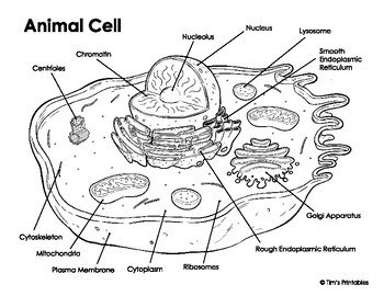
Cell membrane is made up of lipids and proteins and forms a barrier between the extracellular liquid. As observed in the labeled animal cell diagram, the cell membrane forms the confining factor of the cell, that is it envelopes the cell constituents together and gives the cell its shape, form, and existence. This cool worksheet prompts young biologists to research the functions of cell organelles labeled in the diagram. Read more about animal cell, functions and structure of animal. Have cell walls and chloroplasts in contrast to animal cells which have no cell wall or chloroplasts.
Pin on sr1 from i.pinimg.com Cellular water levels biological vector illustration diagram with animal and plant cell. If so, you may need to memorize the animal cell, its organelles, and their functions. Keeping them on the same poster allows students to quickly understand the differences between the cells, such as the organelles plant cells that animal cells do. That cells can be of different shapes and sizes. Phase diagrams the phasediagrams (figure 1a,b,c,d,e)were determined bymaking five different mixturesofpeg anddextran for each ofthe molecularweight. Plant cell and animal cell fall under eukaryotic type. Structure of a eukaryotic cell. Cell membrane is made up of lipids and proteins and forms a barrier between the extracellular liquid.
The animal cell diagram on the free worksheet will teach students to identify the function of the major parts of the animal cell.
The most important structures of plant and animal cells are shown in the diagrams below, which provide a clear illustration of how much these cells have in common. Cell membrane is made up of lipids and proteins and forms a barrier between the extracellular liquid. Read more about animal cell, functions and structure of animal. As observed in the labeled animal cell diagram, the cell membrane forms the confining factor of the cell, that is it envelopes the cell constituents together and gives the cell its shape, form, and existence. Each cell can be thought of as a large phospholipids are molecules with a phosphate group head attached to glycerol and two fatty acid tails. Under the microscope, an animal cell shows many different parts called organelles, that work together to keep the cell functional. The role and function of the plasma membrane; Structure of a eukaryotic cell. During animal cell division, the centrioles replicate (make new copies) and the centrosome divides. Some students may be able to identify some of the structures. Plant cells vs animal cells with diagrams. Printable animal cell diagram to help you learn the organelles in an animal cell in preparation for your test or quiz. Animal cell structure stock illustration.
An animal cell ranges in size from 10 to 30 µm. Read more about animal cell, functions and structure of animal. The animal cell is made up of several structural organelles enclosed in the plasma membrane, that enable it to function properly, eliciting mechanisms that benefit the host (animal). · an animal cell diagram is a great way to learn and understand the many functions of an animal cell. Animal cell drawing at getdrawings.
insotnami: animal cells diagram from 1.bp.blogspot.com These are in the title animal cell parts and functions, the word part pertains to organelles; Unlike the eukaryotic cells of plants and fungi, animal cells do not have a cell wall. Animal cell anatomy diagram structure with all parts nucleus smo. That cells can be of different shapes and sizes. Plant cell and animal cell fall under eukaryotic type. Read more about animal cell, functions and structure of animal. Cells are the basic units of structure and function in living things. They believed that the plasma membrane around cells was made up from a phospholipid bilayer.
Unlike the eukaryotic cells of plants and fungi, animal cells do not have a cell wall.
Each centriole is a ring of nine groups of fused microtubules. Lets us discuss the animal cell, types of an animal cell, animal cell diagram, its structure. During animal cell division, the centrioles replicate (make new copies) and the centrosome divides. Plant vs animal cell diagram in 2020. Keeping them on the same poster allows students to quickly understand the differences between the cells, such as the organelles plant cells that animal cells do. Cytoplasm, ribosomes, rough endoplasmic reticulum; Cellular water levels biological vector illustration diagram with animal and plant cell. If so, you may need to memorize the animal cell, its organelles, and their functions. They believed that the plasma membrane around cells was made up from a phospholipid bilayer. Printable animal cell diagram u2013 labeled unlabeled and blank. The most important structures of plant and animal cells are shown in the diagrams below, which provide a clear illustration of how much these cells have in common. Smooth endoplasmic reticulum, mitochondria, golgi bodies, lysosomes. A labeled diagram of the animal cell and its organelles.


Post a Comment